Showing posts with label nuclear medicine. Show all posts

Tuesday, February 10, 2015

What is a Hepatobiliary Scan

What is a Hepatobiliary Scan? - From the desk of Marcus R Hall


A Hepatobiliary Scan is a Nuclear Medicine Procedure designed to evaluate the patency of the hepatobiliary system's duct work. In other words it checks for blockages, and the functionality of the Gall Bladder.

The blockages can be obstructive objects (gall stones) or infectious processes (inflammations and/or infections).

Hepatobiliary Scans have historically been called HIDA Scan, named for the radiopharmaceutical (hydroxy iminodiacetic acid) used in the earlier exams.

There are additional radiopharmaceeuticals that have been developed over the years, the have been proven to be superior to the original imaging agent. These imaging agents are PIPIDA (paraisopropyl iminodiacetic acid) and DISIDA (disopropyl iminodiacetic acid).


What is a Hepatobiliary Scan - What the patient can expect.


The patient will generally fast overnight as a preparation for the exam, some facilities will allow you to eat at least 4 hours prior to the exam, and has the patient hold any interfering medications to the scan also.

The patient will be placed in the Nuclear Medicine Gamma Camera, where they will receive an injection in an accessible vein; usually the antecubital vein in their arm.

Imaging may start prior to the injection to capture the blood flow to the liver and hepatobiliary system.

The Liver will filter the injection from the blood stream, and send it into the biliary system.

Imaging will continue as this process takes place.

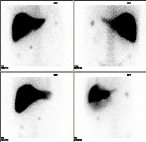
The liver should be the first thing to appear upon injection.

The gall bladder should visualize within an hour.

Following the gall bladder visualization the technologist will continue imaging until the intestinal tract appears.

When the liver, gall bladder, and intestinal tract appear; the procedure is considered complete.

If the intestinal tract hasn't shown up by 2 hours post the injection, the technologist may send the patient to eat. The patient will return for a quick image after eating. If the intestinal tract shows the study is done. If the patient's intestines still haven't shown the technologist will consult the radiologist, and possibly will obtain a 4 hour post injection image.

Following the completion of the exam the radiologist will interpret and transcribe a report for the ordering physician.



Friday, January 23, 2015

Nuclear Medicine Exams For The Layman - Nuclear Medicine Liver/Spleen Scans - From the desk of Marcus R Hall


Nuclear Medicine Exams For The Layman - Nuclear Medicine Liver/Spleen Scans

What is Nuclear Medicine Imaging?

From The Desk Of Marcus R Hall


Nuclear Medicine Live Spleen Scan are done (but not limited to) to


1. Assessing the size, shape, and position of the liver and spleen.

2. Detecting, measuring, and monitoring masses of the liver and/or spleen.

3. Differentiating hepatic hemangiomas (kind of like a blood blister within the liver, have you ever hit your thumb and had a blood blister; but, within the liver)  and focal nodular hyperplasia (a benign tumor) from other liver lesions or tumors.

4. Evaluating how well your liver and spleen are functioning.

5. Confirming the patency of hepatic arterial perfusion catheter. (whether a catheter inserted into the hepatic artery is open, and will allow an injection to be successful made through it).

When you arrive at the imaging center or hospital; you will go to the Nuclear Medicine Department.

If the facility wants to perform a Liver Blood Flow; you will be taken to lie done on the Nuclear Medicine Imaging Table, and placed under the Nuclear Medicine Camera.

The Nuclear Medicine Technologist will administer a radiopharmaceutical injection of technitium99m bound to sulfide particles; otherwise know as technetium sulfur colloid.

As the technetium starts to decay, it releases gamma rays or light particles, at the sub-atomic level; these particles are called photons. Your naked eye cannot see these particles; the Nuclear Medicine can.

What the Liver Blood Flow allows the Nuclear Radiologist to evaluate the blood flow to the Liver and Spleen.

Following your injection it takes 15 or 20 minutes to reach a peak accumulation in the Liver and the Spleen.

At approximately 15 to 20 minutes the Liver Imaging will be done.

There are two basic imaging methods, planar or static imaging and SPECT imaging or a combination of both.

Planar imaging or static imaging is an image that is taken in one position at one time.
The typical images are the anterior position (in front of you), Right Anterior Oblique (right front, at an angle), Right Lateral (your right side), Right Posterior Oblique (right back side at an angle), Posterior (straight from the back), Left Posterior Oblique (left back side), Left Lateral (left side) and Left Anterior Oblique (left front side at an angle).

SPECT Imaging is Single Photon Emission Computed Tomography. The Nuclear Takes images 360 degrees around your body, and the computer can reconstruct the images into a rotating 3D image of your liver and spleen allowing localization of hemangiomas, and other lesions.

If you're being evaluated for a hemangioma they generally will use red blood cells (drawn from your arm, and re injected once tagged with a radioisotope) and image when you are re injected with your blood. twenty minutes after your re injection, and two to three hours after your re injection of the tagged red blood cells. So, you will have multiple visits to have your scan completed.

This information is not medical advise; this must come from your Doctor. This is imformation so a patient isn't going blind into a procedure.  It is intended to assist the patient in planning the exam day.

To Your Health - Marcus R Hall.

Tuesday, December 2, 2014

Nuclear Medicine Three Phase Bone Scans a.k.a. Tri-Phase Bone Scans

Nuclear Medicine Three Phase Bone Scans a.k.a. Tri-Phase Bone Scans


From The Desk Of Marcus R Hall

Nuclear Medicine Three Phase Bone Scans are done primarily to detect bone infections, stress fractures, and evaluate pathological processes involving the bone.

A Three Phase Bone Scan is done in a similar fashion as a Bone Scan with exception to imaging being done at three times during the progression of the study.

The Blood Flow Portion of the study. Imaging is done as the radiopharmaceutical is administered intravenously to the patient.  This is done to visualize and evaluate a balanced blood through the area of interest.  Generally, this involves the extremities of the patient i.e., blood flow to the feet or hands.

Three Phase Bone Blood Flow

The Blood Flow a.k.a. Bone Flow, is generally completed within 2  minutes of  receiving the administration of the radiopharmaceutical. The blood flow portion of the exam is not interpreted until the entire study is completed.

The Blood Pool portion of the Three Phase Bone Scan follows the Blood Flow portion of the scan.

The Blood Pool portion of the scan is to document redness and swelling in the area of interest. Images are taken generally 10 - 15 minutes after the administration of the radiopharmaceutical.

Nuclear Medicine Blood Pool Images


Following the Blood Pool portion of the scan; the patient is generally released for a few hours, and asked to force fluids to flush the soft tissues, so just bones are visible on their return.

On the patients return images are taken of the area of interest; this is generally a thirty minute imaging session.  Don't become alarmed if the technologist takes images at multiple angles or views.
Nuclear Medicine Delay Bone Images

Upon completion of the study, a Physician trained in interpreting Nuclear Medicine Procedures will report to your requesting Physician the result of the scan.

The purpose of this blog post is purely informational for patients; it is not meant to serve as medical advice. This is just a short informational blurb to let patients have an understanding as to what they may experience in the performance of their exam. There may be slight variations in the procedure between facilities. These variations don't make one facility right, nor one wrong; just different...yielding the same results.

My Doctor Has Ordered A Nuclear Medicine Bone Scan - Why and What For?

My Doctor Has Ordered A Nuclear Medicine Bone Scan - Why and What For?

From The Desk Of Marcus R Hall

Nuclear Medicine Bone Scans are done to determine bone damage due to physical or pathological trauma.


Nuclear Medicine Bone Scans allow Clinicians to image the actual physiological function of the bone, and document it with images and statistical data stored on a computer or films.

The skeletal system is always in a constant state of repair replacing dead or dying bone cells to maintain the skeleton.  This repair uses crystals on the surface of the bone where normal maintenance is taking place, in areas of bone damage the crystals are more concentrated.

Clinicians can "tag" or attach a radioactive pharmaceutical to these pre-bone crystals. The radioactive isotope in the pharmaceutical releases energy that they can be seen and imaged using a special Nuclear Medicine Camera.

The Nuclear Medicine Bone Scan Begins With A Radioactive Injection.

The procedure begins with the patient reporting to a Nuclear Medicine Department licensed to perform these exams, with specially trained Physician's who interpret the exams.

The patient receives an intravenous injection of a radioactive phosphate that attaches itself to the patients pre-bone repair crystals.  

In order to see the bones approximately three hours later ... it is necessary to flush the remaining injection circulating through the circulatory system, soft tissue, muscles, and skin. 

The patient will be asked to force fluids, utilizing the patent's  kidneys to flush out the excess injection.

Following the flushing out period which can vary between facilities dependent on the exact radiopharmaceutical administered to the patient. The patient will return to the facility for the imaging portion of the study.

Imaging takes place with a Nuclear Medicine Camera, which can see and record the flashes of energy released by the bone scan injection; which is now attached to the crystals that are repairing the bone.
Photo Compliments of Wikipedia


The imaging process may be of an individual area of interest, or may include the entire body.
Don't get too concerned if the Technologist takes images at different angles of positions... it doesn't necessarily mean there is anything wrong.  To do the exam correctly, they just need to be thorough.

A Physician trained in Nuclear Medicine will interpret your exam results, and report them back to your requesting Physician.


The purpose of this blog post is purely informational for patients; it is not meant to serve as medical advice. This is just a short informational blurb to let patients have an understanding as to what they may experience in the performance of their exam. There may be slight variations in the procedure between facilities. These variations don't make one facility right, nor one wrong; just different...yielding the same results.