Nuclear Medicine Exams For The Layman - Nuclear Medicine Liver/Spleen Scans
What is Nuclear Medicine Imaging?
From The Desk Of Marcus R Hall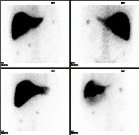
Nuclear Medicine Live Spleen Scan are done (but not limited to) to
1. Assessing the size, shape, and position of the liver and spleen.
2. Detecting, measuring, and monitoring masses of the liver and/or spleen.
3. Differentiating hepatic hemangiomas (kind of like a blood blister within the liver, have you ever hit your thumb and had a blood blister; but, within the liver) and focal nodular hyperplasia (a benign tumor) from other liver lesions or tumors.
4. Evaluating how well your liver and spleen are functioning.
5. Confirming the patency of hepatic arterial perfusion catheter. (whether a catheter inserted into the hepatic artery is open, and will allow an injection to be successful made through it).
When you arrive at the imaging center or hospital; you will go to the Nuclear Medicine Department.
If the facility wants to perform a Liver Blood Flow; you will be taken to lie done on the Nuclear Medicine Imaging Table, and placed under the Nuclear Medicine Camera.
The Nuclear Medicine Technologist will administer a radiopharmaceutical injection of technitium99m bound to sulfide particles; otherwise know as technetium sulfur colloid.
As the technetium starts to decay, it releases gamma rays or light particles, at the sub-atomic level; these particles are called photons. Your naked eye cannot see these particles; the Nuclear Medicine can.
What the Liver Blood Flow allows the Nuclear Radiologist to evaluate the blood flow to the Liver and Spleen.
Following your injection it takes 15 or 20 minutes to reach a peak accumulation in the Liver and the Spleen.
At approximately 15 to 20 minutes the Liver Imaging will be done.
There are two basic imaging methods, planar or static imaging and SPECT imaging or a combination of both.
Planar imaging or static imaging is an image that is taken in one position at one time.
The typical images are the anterior position (in front of you), Right Anterior Oblique (right front, at an angle), Right Lateral (your right side), Right Posterior Oblique (right back side at an angle), Posterior (straight from the back), Left Posterior Oblique (left back side), Left Lateral (left side) and Left Anterior Oblique (left front side at an angle).
SPECT Imaging is Single Photon Emission Computed Tomography. The Nuclear Takes images 360 degrees around your body, and the computer can reconstruct the images into a rotating 3D image of your liver and spleen allowing localization of hemangiomas, and other lesions.
If you're being evaluated for a hemangioma they generally will use red blood cells (drawn from your arm, and re injected once tagged with a radioisotope) and image when you are re injected with your blood. twenty minutes after your re injection, and two to three hours after your re injection of the tagged red blood cells. So, you will have multiple visits to have your scan completed.
This information is not medical advise; this must come from your Doctor. This is imformation so a patient isn't going blind into a procedure. It is intended to assist the patient in planning the exam day.
To Your Health - Marcus R Hall.