Tuesday, February 10, 2015

What is a Hepatobiliary Scan

What is a Hepatobiliary Scan? - From the desk of Marcus R Hall


A Hepatobiliary Scan is a Nuclear Medicine Procedure designed to evaluate the patency of the hepatobiliary system's duct work. In other words it checks for blockages, and the functionality of the Gall Bladder.

The blockages can be obstructive objects (gall stones) or infectious processes (inflammations and/or infections).

Hepatobiliary Scans have historically been called HIDA Scan, named for the radiopharmaceutical (hydroxy iminodiacetic acid) used in the earlier exams.

There are additional radiopharmaceeuticals that have been developed over the years, the have been proven to be superior to the original imaging agent. These imaging agents are PIPIDA (paraisopropyl iminodiacetic acid) and DISIDA (disopropyl iminodiacetic acid).


What is a Hepatobiliary Scan - What the patient can expect.


The patient will generally fast overnight as a preparation for the exam, some facilities will allow you to eat at least 4 hours prior to the exam, and has the patient hold any interfering medications to the scan also.

The patient will be placed in the Nuclear Medicine Gamma Camera, where they will receive an injection in an accessible vein; usually the antecubital vein in their arm.

Imaging may start prior to the injection to capture the blood flow to the liver and hepatobiliary system.

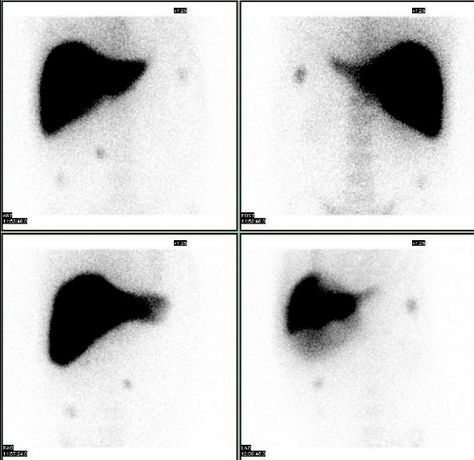
The Liver will filter the injection from the blood stream, and send it into the biliary system.

Imaging will continue as this process takes place.

The liver should be the first thing to appear upon injection.

The gall bladder should visualize within an hour.

Following the gall bladder visualization the technologist will continue imaging until the intestinal tract appears.

When the liver, gall bladder, and intestinal tract appear; the procedure is considered complete.

If the intestinal tract hasn't shown up by 2 hours post the injection, the technologist may send the patient to eat. The patient will return for a quick image after eating. If the intestinal tract shows the study is done. If the patient's intestines still haven't shown the technologist will consult the radiologist, and possibly will obtain a 4 hour post injection image.

Following the completion of the exam the radiologist will interpret and transcribe a report for the ordering physician.


Saturday, February 7, 2015

What Is A VQ Scan For Lungs? - Information For The Layman

What is a VQ Scan for Lungs a.k.a. Lung VQ, Lung VP.

From the desk of Marcus R Hall - Retired Nuclear Medicine Technologist

A VQ scan for the lungs is a Nuclear Medicine Procedure whose purpose is to detect and diagnose a blood clot in the lungs, evaluating chronic vs. acute PE, and or staging COPD.
                                                              VP Comparison Depicting PE

Some of the clinical indicators to warrant a Nuclear Medicine Lung VQ Scan include:
  • shortness of breath (developed after a long period of being stationary ie; long plane, train, train, or automobile trips.
  • a history of COPD (asthma, emphysema)
  • a history of pulmonary emboli (blood clots in the lungs).
  • a drop in oxygen saturation levels following surgery.
What to expect.
The patient reports to the imaging center. The technologist will explain the above information to the patient. Techniques and protocols may vary between imaging centers; but, will be similar with the same data collection at the end.  This data may be manipulated by the Nuclear Medicine Technologist based on the Physicians Orders, Suspected Diagnosis, etc.

Phase One The Ventilation Portion (V) of the exam may be done with a radioactive Xenon Gas which the patient breathes in and out while images are taken with a Nuclear Medicine Camera. This data may be used to determine wash in, equilibrium, and washout timing images as well as any obstructions of the lungs.

A DTPA-Tc99m aerosol may be used for the ventilation, it depends on which protocol the facility is licensed to perform, and equipment on hand.
A Xenon Washin-Equalibrium-Wash Out
Ventilation Image                                                               A Tc99m-DTPA Ventilation Image

Phase Two - Quantification and/or Perfusion Portion of the Exam.

A Tc99m-DTPA Perfusion Image


In a Quantification Study the data collected is manipulated to determine the sectional function and perfusion of the lungs for patient's facing the removal of a portion or an entire lung. 

In a Perfusion Study the Technologist will acquire and manipulate the data to evaluate for a pulmonary emboli, a.k.a.

For the Perfusion Portion of the Nuclear Medicine VQ scan the patient is injected with a radioactive pharmaceutical that localizes in the lungs relative to blood flow.

Blood clots in the lungs are evident as segmental or sub-segmental defects in the lung images as shown in the image below.


The Patient will lay or sit in the Nuclear Medicine camera while images are taken of the lungs.

The entire procedure will take as long as 30 minutes to one hour, dependent on the patients condition.

Upon completion, the Radiologist will interpret the results of the exam, and report them to the requesting Physician.


Friday, January 23, 2015

Nuclear Medicine Exams For The Layman - Nuclear Medicine Liver/Spleen Scans - From the desk of Marcus R Hall


Nuclear Medicine Exams For The Layman - Nuclear Medicine Liver/Spleen Scans

What is Nuclear Medicine Imaging?

From The Desk Of Marcus R Hall


Nuclear Medicine Live Spleen Scan are done (but not limited to) to


1. Assessing the size, shape, and position of the liver and spleen.

2. Detecting, measuring, and monitoring masses of the liver and/or spleen.

3. Differentiating hepatic hemangiomas (kind of like a blood blister within the liver, have you ever hit your thumb and had a blood blister; but, within the liver)  and focal nodular hyperplasia (a benign tumor) from other liver lesions or tumors.

4. Evaluating how well your liver and spleen are functioning.

5. Confirming the patency of hepatic arterial perfusion catheter. (whether a catheter inserted into the hepatic artery is open, and will allow an injection to be successful made through it).

When you arrive at the imaging center or hospital; you will go to the Nuclear Medicine Department.

If the facility wants to perform a Liver Blood Flow; you will be taken to lie done on the Nuclear Medicine Imaging Table, and placed under the Nuclear Medicine Camera.

The Nuclear Medicine Technologist will administer a radiopharmaceutical injection of technitium99m bound to sulfide particles; otherwise know as technetium sulfur colloid.

As the technetium starts to decay, it releases gamma rays or light particles, at the sub-atomic level; these particles are called photons. Your naked eye cannot see these particles; the Nuclear Medicine can.

What the Liver Blood Flow allows the Nuclear Radiologist to evaluate the blood flow to the Liver and Spleen.

Following your injection it takes 15 or 20 minutes to reach a peak accumulation in the Liver and the Spleen.

At approximately 15 to 20 minutes the Liver Imaging will be done.

There are two basic imaging methods, planar or static imaging and SPECT imaging or a combination of both.

Planar imaging or static imaging is an image that is taken in one position at one time.
The typical images are the anterior position (in front of you), Right Anterior Oblique (right front, at an angle), Right Lateral (your right side), Right Posterior Oblique (right back side at an angle), Posterior (straight from the back), Left Posterior Oblique (left back side), Left Lateral (left side) and Left Anterior Oblique (left front side at an angle).

SPECT Imaging is Single Photon Emission Computed Tomography. The Nuclear Takes images 360 degrees around your body, and the computer can reconstruct the images into a rotating 3D image of your liver and spleen allowing localization of hemangiomas, and other lesions.

If you're being evaluated for a hemangioma they generally will use red blood cells (drawn from your arm, and re injected once tagged with a radioisotope) and image when you are re injected with your blood. twenty minutes after your re injection, and two to three hours after your re injection of the tagged red blood cells. So, you will have multiple visits to have your scan completed.

This information is not medical advise; this must come from your Doctor. This is imformation so a patient isn't going blind into a procedure.  It is intended to assist the patient in planning the exam day.

To Your Health - Marcus R Hall.